
A revolution in Neuroanatomy!
There has been a revolution in the unlikeliest of disciplines! Neuroanatomy!
Yes neuroanatomy – so cut and dry – has been seriously altered by new research. The textbook world of neuroanatomy turned upside down yesterday. A team led by Jean François Brunet of INSERM in Paris has made the audacious proposal that the autonomic outflow of the sacral cord is sympathetic rather than parasympathetic.
Unfortunately, this article is not freely available. I was able to read the entire article because I am associated with a university. Open access is a big problem, particularly at a time when the public is inundated with so much scientifically questionable information. If the scientific community wants to facilitate the public’s scientific literacy, achieving universal open access would be a good place to start. But I digress….
When I heard the news – sacral autonomic outflow is sympathetic not parasympathetic – the image that kept flashing through my mind’s eyes was the Roy Lichtenstein cartoon where a woman laying languidly in bed looks up with mild alarm and says, “Oh my God! I left the baby on the bus!” Well, if the results by Brunet’s group, led by first author Isabel Espinosa-Medina, hold up, I can imagine an anthropomorphized parasympathetic system, “Parasympathia,” saying:
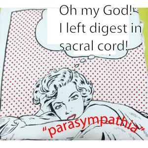
Fill in your own bubble words here in this take-off from Roy Lichtenstein’s iconic cartoon.
The approach
Espinosa-Medina, Brunet and colleagues approached this question using a now-common modern molecular approach. The approach goes something like this:
- Find lots of molecules that are present in Structure A
- Find a non-overlapping set of molecules present in Structure B
- Determine if Structure C has Structure A- or Structure B-associated molecules
My colleague Cliff Ragsdale, working with then graduate students Jennifer Dugas-Ford and Joanna Rowell, used this to show that bird telencephalon is organized in a homologous manner to mammalian neocortex. As you may know, only mammals have a 6-layered neocortex. But Harvey Karten proposed, many years ago, that the bird brain has areas that are homologous to neocortical layers. Cliff’s laboratory examined this idea by identifying non-overlapping sets of molecules that reliably mark either the input (layer 4) or output (layer 5) layers of mammalian neocortex. Then they looked in the bird brain. The molecules that mark input layer 4 (e.g. EAG2, RORB) are also found in specific nuclei (e.g. entopallium) of the bird brain whereas molecules that mark output layer 5 of mammals (e.g. ER81, FEZF2) are found in different regions of the bird brain (e.g.arcopallium). These results tell us that while the architecture of bird and mammal brains differs, the development mechanisms and evolutionary history are at least partially shared. Moreover, to use computer science language, the results hint at similarities between the processing capacities of the bird “telencephalic chip” and the “neocortical chip.”
The evidence
And now back to the sacral autonomic outflow and the findings of Brunet’s laboratory. Espinosa-Medina and her colleagues found non-overlapping sets of molecular markers and characteristics for:
- Preganglionic autonomic neurons in thoracic cord – these are for sure sympathetics
- Preganglionic autonomic neurons in the hindbrain-located dorsal motor nucleus of the vagus (dMX) – these are for sure parasympathetics (it doesn’t get any more parasympathetic than the vagus…)
They then used the markers to examine the autonomic neurons in the sacral cord. Lo and behold, they found that the sacral autonomic motor neurons had the same characteristics as the thoracic preganglionics (jargon for thoracic preganglionic neurons; excuse the ungrammatical adjective-as-noun) and a wholly different set of features than the cranial preganglionics. The Brunet laboratory employed a number of molecular markers with alphabet-soup-names such Phox2B, Foxp1, Tbx, and VAChT. The markers aligned as predicted by a parasympathetic identity for the sacral autonomic motor neurons (aka preganglionics).
One particularly compelling experiment was testing the nerve-dependence of sacral ganglionic development. As it turns out (I just learned this myself), sympathetic ganglia develop independently of an innervating nerve whereas parasympathetic ganglia (in the head) require the in-growth of innervating nerve fibers. In a mouse that was genetically engineered to lack sacral nerve outgrowth, the pelvic ganglion still formed. This means that the ganglion innervated by sacral cord has a developmental characteristic that aligns with the sympathetic rather than the parasympathetic system.
Potential limitations
Overall, I really like this work and I am largely convinced. Just one major issue worries me.
My primary concern is the omission from consideration of the parasympathetic preganglionics that exit from the midbrain through cranial nerve III (aka oculomotor). The axons of these autonomic preganglionics have a ventral exit point as do the sympathetic preganglionics from thoracic (and now sacral) cord. This is in contrast to the dorsolateral exit point for parasympathetics (such as the vagus) that exit from the hindbrain. In addition, the ciliary ganglion (which receives innervation from autonomic nerve fibers in the oculomotor nerve) expresses Islet1, a marker of sympathetic ganglia. A very cursory search on mechanisms of ciliary ganglion development reveals additional ways in which the ciliary ganglion resembles sympathetic ganglia. There may be more.
Trying to fit the autonomic outflow from the midbrain into the new classification muddies the picture. Accepting the results of Brunet and colleagues, I see three possibilities:
- Parasympathetics emanate from the midbrain and hindbrain whereas sympathetics emanate from the spinal cord (autonomic CN III fibers are parasympathetics)
- Parasympathetics emanate from the hindbrain only while sympathetics emanate from the spinal cord and midbrain (autonomic CN III fibers are sympathetics)
- Parasympathetics emanate from the hindbrain, sympathetics emanate from the spinal cord and the jury is out on how to accurately classify the midbrain autonomic outflow (autonomic CN III fibers may be in their own distinct category)
I am guessing that # 1 is correct (as Brunet and colleagues would argue) but I would not rule out #2 or #3.
Implications
Chances are that the authors are correct in concluding that thoracic and sacral autonomic neurons belong to the same sympathetic division. I will divide the potential implications into three categories:
What are the consequences for teaching?
The teaching implications are major. We have to change what we teach for a few reasons. First, we want to teach truths. Once we know something is wrong, it is incumbent upon us to stop teaching an untruth. Typically, during the time that is required for a new truth to be accepted, I would argue that teaching what used to be thought along side with the new truth is warranted. This enables students to understand confusions that can arise when they are talking with someone who has not yet been made aware of the new findings. [Additionally it is non-trivial that students need to pass national examinations that are based on information that lags [probably years] behind the most recent findings.]
How important is this finding to our understanding of the nervous system?
Here, the finding is very important because it places the sacral autonomics squarely into the developmental and evolutionary category of the thoracic sympathetics. What I mean by this is that the sacral and thoracic autonomic outflows develop by similar mechanisms and share a common evolutionary history. A common evolutionary history suggests common selection pressures which is evidence (albeit indirect) for common functions of thoracic and sacral autonomic outflows.
What are the functional implications?
I suspect that robust discussions that occur over some time will be necessary to fully work out the functional implications of the new paradigm. However, Brunet and colleagues point out that the common categorical identity of thoracic and sacral autonomics lend support to cooperative over antagonistic interactions between the two areas.
Presently highly distinct functions are ascribed to the thoracic (sympathetic) and sacral (heretofore parasympathetic) innervations of the bladder and sex organs. In the case of the bladder, the thoracic innervation is thought to modulate elasticity of the bladder, thereby changing the volume at which a sense of fullness occurs, and the sacral innervation is thought to lead to bladder contraction and thus voiding. In the case of the sex organs, thoracic outflow is critical to sexual arousal while sacral outflow is critical to sexual climax. Clearly, in both cases, the two regions work together and not antagonistically to achieve the desired end. Placing thoracic and sacral autonomic outflows in the same category has the advantage of semantically emphasizing the synergy of the two regions.
A historical view of the autonomic system antagonism
I want to end this post by commenting on a quote from Walter Gaskell who initially described the parasympathetic and sympathetic divisions of the autonomic nervous system in the late 1800s. In 1886, Gaskell wrote:
“The evidence is becoming daily stronger that every tissue is innervated by two sets of nerve fibers of opposite characters so that I look forward hopefully to the time when the whole nervous system shall be mapped into two great districts of which the function of the one is katabolic [breaking down], of the other anabolic [building up], to the peripheral tissues: two great divisions of the nervous system which are occupied with … opposite chemical processes.”
Gaskell sets up a battle between the forces of tear-down and those of build-up. It hardly takes imagination to think of re-purposing this relationship as one between good and evil. And it is this antagonism that has made its way into the minds and memories of the public. Even school children have heard of the dichotomy between fight-or-flight and rest-and-digest. The implied common evolutionary heritage of thoracic and sacral outflows emphasizes that one extreme function is not useful without the other. Furthermore, our current understanding of autonomic control emphasizes balanced and target-specific regulation over massive excitement of either autonomic division.
In the end, I suspect that the roots of the antagonistic picture of sympathetic and parasympathetic function are more historical than biologically accurate. This is just a guess and a prod toward a historian out there looking for a new project.
On a personal note
In an odd coincidence, the Brunet article came out on the day that I finished correcting the page proofs for the 2nd edition of my textbook, Medical Neurobiology. I feel this coincidence as the nervous system’s mocking my hubris that I could capture a tell-no-lies version of neuroanatomy. Clearly, my telling bears an un-truth within before it is even published. Sadness.
Neuroanatomy does not change from one year to another or one book edition to the next. But our understanding of neuroanatomy changes all the time. Usually our scientific knowledge moves forward step by step but happily we just experienced a leap forward.
Categories: Brain anatomy, Brain Function


If Espinosa-Medina, Brunet and colleagues are proven correct I wonder if any ex students will be asking for a re-marking … 😆
It is good to learn that advances continue tho I profoundly agree that access to papers should not mirror the medieval monks hegemony over ‘the truth’.
LikeLike
I think the new Coursera.org also has the old info..”who knew?”
LikeLike
Indeed. The new info has been publicly available for three days, actually less than 72 hours. So no existing document or video had those information incorporated. I wonder if Wikipedia is up to date.
LikeLike
Just checked Wikipedia and no, not updated. That may be appropriate until there is some independent confirmation.
LikeLike
… Thank you for this post. It reminds me again that all knowledge is temporary until proven wrong. And me personally I like the view that all the elements in our body work together without central control in some way or another, like ‘thoracic and sacral autonomics’. Only e.g. chronic stress produces a breach of this cooporation of different talents which leads to ‘disease’. …
LikeLike
I agree. Everything has truth until it doesn’t.
After more reflection, I’ve come to think that the take home message is not that the sympathetic-parasympathetic dichotomy doesn’t exist. It does. And it still is well summarized in the fight or flight vs rest and digest vernacular. BUT the new bit of that voiding both urine and feces and also sexual arousal and climax are part of the flight or fight, sympathetic system.
LikeLike
Are we forgetting the postganglionics here? Presumably the postganglionics innervated by the sacral outflow can be considered sympathetic cholinergic and hence can still be ‘antagonistic’ to the ‘classic’ sympathetics
LikeLike